


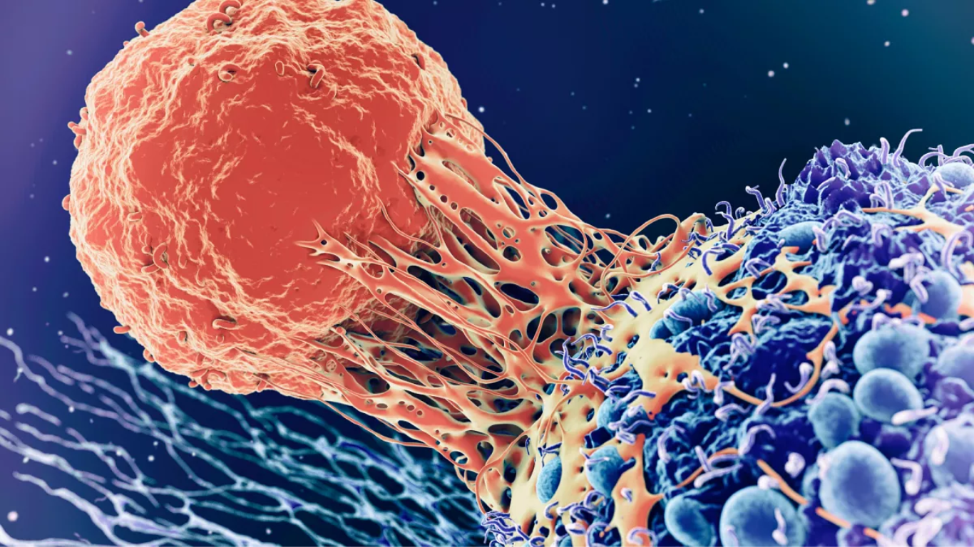


According to a group of neuroscientists, the size of our primary visual cortex and the quantity of brain tissue dedicated to processing visual information at certain points in visual space might predict how well we see. The findings point to a new link between brain architecture and behaviour.
The findings of the study were published in the journal ‘Nature Communications.’ “We discovered that we can predict how well someone can see based on the distinctive form of their primary visual cortex,” says lead author Marc Himmelberg, a postdoctoral researcher at New York University’s Center for Neural Science and Department of Psychology.
“We can better understand what underpins variations in how people perceive and interact with their visual surroundings by demonstrating that individual diversity in the anatomy of the human visual cortex is connected to variance in visual functioning.”
The bumps and grooves on each person’s brain surface, like fingerprints, are unique. The importance of these variations, however, is not entirely known, particularly when it comes to their influence on behaviour, such as disparities in our capacity to perceive.
Himmelberg and his co-authors, NYU’s Center for Neural Science and Department of Psychology professors Jonathan Winawer and Marisa Carrasco, aimed to elucidate the importance of these brain features to how we perceive in the Nature Communications study.
The primary visual cortex (V1) organises a picture projected from the eye into a map. However, it is distorted, as are many maps, with some areas of the image being larger than others.
“Think of a subway map of New York City that makes Staten Island appear smaller than Manhattan,” Winawer adds. “The map retains some accuracy, but it enlarges locations that are likely to be of greater interest. Similarly, V1 enlarges the centre of the picture we view, i.e. where our eyes are fixated, in comparison to the periphery.”
This is due to V1 having more tissue devoted to the centre of our field of vision. Similarly, V1 enlarges places to the left and right of where our eyes are fixating in comparison to regions above or below, due to changes in cortical tissue organisation.
The scientists used functional magnetic resonance imaging (fMRI) to map the size of the primary visual cortex (or “V1”) in more than two dozen adults. The amount of V1 tissue these people had allocated to processing visual information from distinct positions in their field of view—locations to the left, right, above, and below fixation—was also assessed.
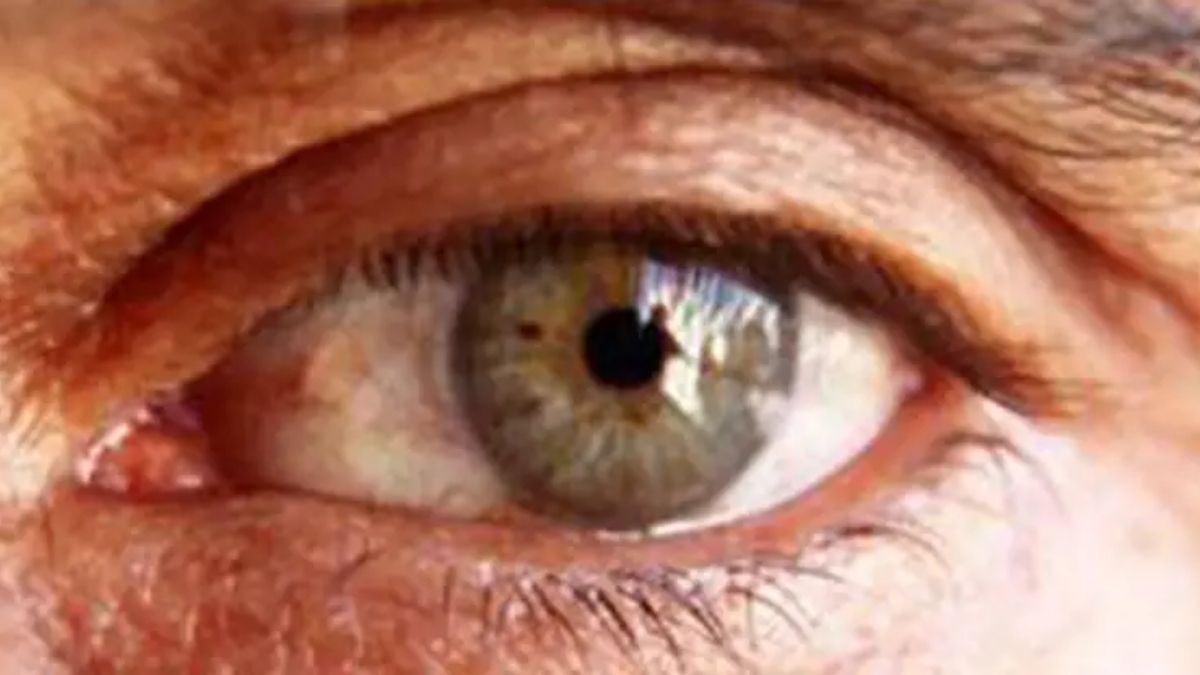
These participants also completed a task designed to evaluate their vision quality at the same areas in their field of view as the V1 assessments. The participants differentiated between the orientations of patterns displayed on a computer screen, which was used to assess “contrast sensitivity,” or the capacity to distinguish between pictures.
Their findings demonstrated that variations in V1 surface area might predict assessments of people’s contrast sensitivity. First, persons with a high V1 showed higher overall contrast sensitivity than those with a low V1 (the largest surface area is 1,776 square millimetres [mm2] and the smallest is 832 mm2).
Second, persons whose V1 had more cortex tissue processing visual input from a specific location in their field of vision had higher levels of cortical tissue.
Second, persons with more cortical tissue dedicated to processing visual information from a given place in their field of vision had better contrast sensitivity in that region than those with less cortical tissue dedicated to the same region. Third, stronger contrast sensitivity at a certain position (e.g., left) compared to another place equidistant from fixation (e.g., above) linked to areas with more or less cortical tissue, respectively, across individuals.
“In summary,” Carrasco writes, “the more local V1 surface area allocated to encoding a given region, the better the vision at that site.” “Our findings reveal that disparities in visual perception are inexorably connected to differences in the brain’s primary visual cortex anatomy.”